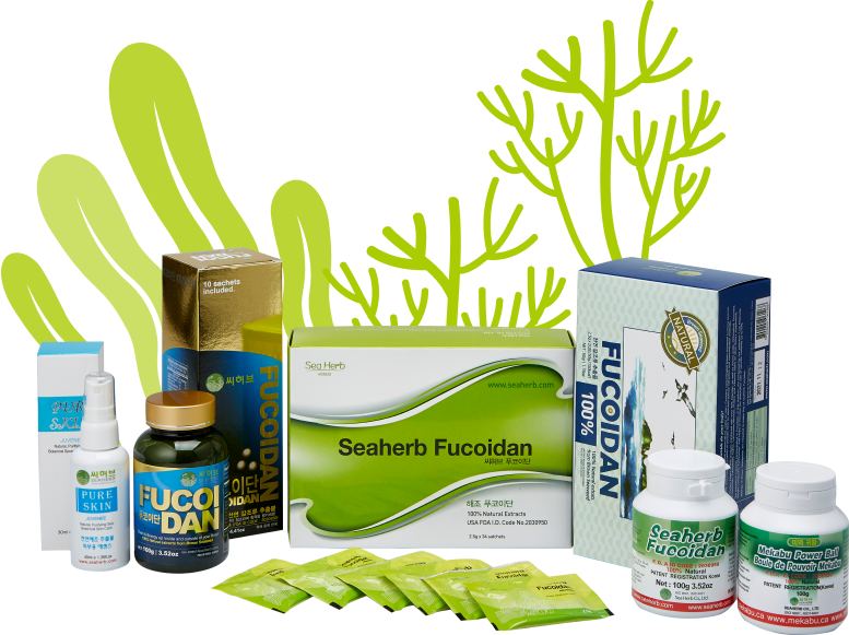
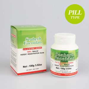
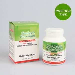
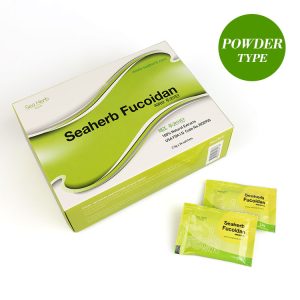
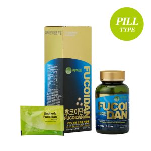
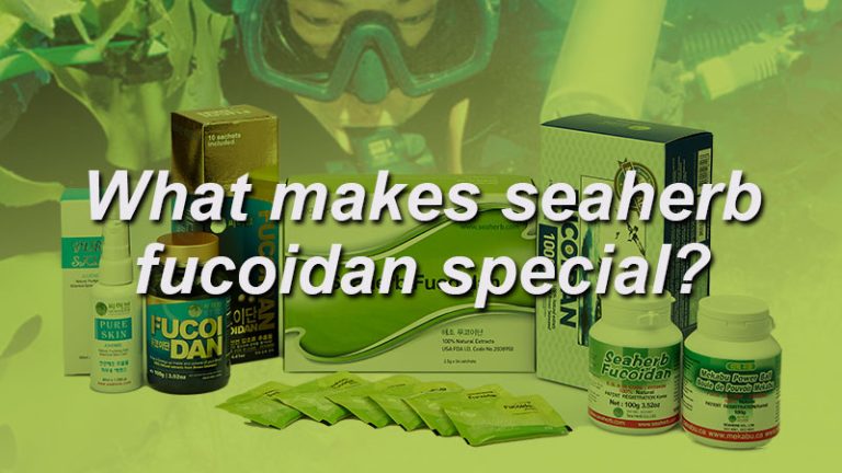
SeaHerb Fucoidan is The 100% natural extract of life-extending substances in this product are enhanced by our patented “Low Molecular Weight Compound Technology” that helps to increase the absorption rate of specific nutrients up to 57%.
BEST ITEM
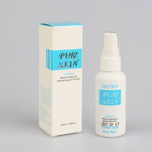
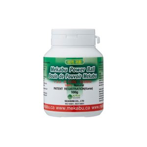
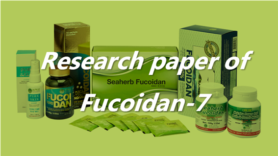
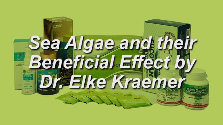
Meet a variety of products from SeaHub’s best items